Rupture Calcaneal Tendon

The calcaneal tendon, also referred to the Achilles tendon (named after the Greek war hero in Homer’s Iliad). The calcaneal tendon is the strongest, thickest, and most powerful tendon in the body. It is an extension of two leg muscles: the gastrocnemius and soleus; the tendon is approximately 15cm long. The strength of the tendon was proved during a stress test, which showed that the tendon could with stand a load of 3.9 times body weight while walking and about twice that when running. The tendon begins at middle of the triceps surae and distally attaches to the posterior surface of the calcaneal tuberosity.
The rupturing of the calcaneal tendon is very traumatic and is often seen in poorly conditioned people. The trauma is typically experienced as an audible snap during a forceful plantarflexion with the knee extended followed by sudden triceps surae pain and dorsiflexion of the plantarflexed foot. The rupturing of the calcaneal tendon is the most serve acute muscular problem of the leg. Ambulation is possible but only when the limb is laterally rotated. A hematoma appears in the malleolar region; along with a lump on the calf owing to the shorting of the triceps surae. Surgical intervention is necessary for athletes and people with active lifestyles, but older and non-athletic people usually are able to rehab without a surgical intervention.
http://en.wikipedia.org/wiki/Achilles_tendon
Hamstring Injuries
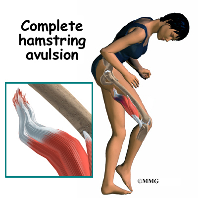
The Hamstring is comprised of three posterior thigh muscles: biceps femoris, semitendinosus, and semimembranosus. The hamstrings cross and act upon two joints: the hip and knee. The semitendinosus and semimembanosus extend the hip when the trunk is fixed; they also flex the knee and medially rotate the lower leg. The biceps femoris extends the hip during walking; short and long heads flex the knee and laterally rotates the lower leg when the knee is bent. The hamstrings play a crucial role in many daily activities like walking, jumping, and controlling some movement in the trunk. The hamstring acts as an antagonist to the quadriceps during most of the activities listed above.
Hamstring trauma, pulled and/or torn, are common in individuals who run and/or kick hard during athletic activities. This trauma is twice as common as quadriceps strains. The violent muscular exertion required to excel in these sports may avulse part of the proximal tendinous attachments of the hamstrings to the ischial tuberosity. Tearing of the hamstrings is a very painful trauma, and are often caused by inadequate warm up and stretching before physical activity. Depending on the servarity of the trauma to the hamstrings the rehab regime is straight forward; us cold therapy and compression bandages during the first 48hrs of injury followed by messaging and stretching the muscle. Only under extreme cercumstances is a surgical intervention necessary.
http://en.wikipedia.org/wiki/Hamstring
Femoral Hernias
A hernia occurs when the contents of the abdomen, usually part of the small intestine, push through a weak point or tear the thin muscular wall of the abdomen, which holds the abdominal organs in place. The femoral rings, the base of the femoral canal, is a weak area in the anterior abdominal wall. It’s a long diameter measures about 1.25cm, and its boarder includes the inguinal ligament, pectineus muscle, lacunar ligament, and the medial side of the femoral vein.
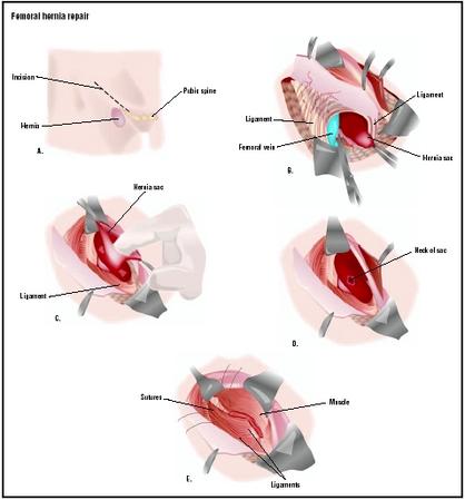
The femoral ring is the usual site for femoral hernias, a protrusion of abdominal viscera through the femoral ring into the femoral canal. The hernia appears as a mass in the femoral triangle, inferolateral to the pubic tubercle. The hernial sac compresses the contents of the femoral canal and distends the wall of the canal. Initially the hernia is small because it is contained within the canal, but it can enlarge by passing inferiorly through the saphenous opening into the subcutaneous tissue of the thigh. Femoral hernias are more common in women because of their wider pelves. Necrosis can occur during strangulation of a femoral hernia because of the sharp, rigid boundaries of the femoral ring, particularly the concave margin of the lacunar ligament. Treatment is usually a surgical intervention, often a piece of plastic mesh is surgically placed to repair the defect in the abdominal wall.
http://en.wikipedia.org/wiki/Femoral_hernia
No comments:
Post a Comment