Mallet/Baseball Finger
 The extensor digitorum tendon at the distal interphalangeal joint is the location of trauma during mallet finger. The extensor digitorum muscle is located in the posterior forearm extending from the lateral epicondyle of the humerus and distally attaching to extensor expansions of medial four digits,. Interphalangeal joints are hinge joints between the phalanges of the hand; the distal interphalangeal joints are those between the second and third phalanges.
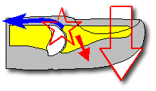 This trauma results from the distal interphalangeal joint suddenly being forced into extreme flexion, for example, while a baseball is miscaught or a finger is jammed into the base pad. Treatment may or may not include a surgical intervention, depending on the severity of the deformity a Mallet splint can be worn for 6 to 8 weeks. The splint allows the tendon to return to normal length on its own. Surgery is used to reattach the tendon and is usually performed within a week of the injury.
Fracture of Olecranon
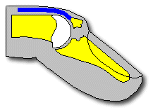
The olecranon process of the ulna is a thick curved bony eminence of the forearm that projects behind the elbow. It is located at the proximal end of the ulna and aids in the hinge joint of the elbow. The olecranon process of the ulna is the distal attachment point for the triceps brachii and the anconeus muscle. Branches of the ulnar nerve are found lying over the olecranon process of the ulna.

http://orthoinfo.aaos.org/topic.cfm?topic=A00503
Injury of Ulnar Nerve at Elbow
 The Ulnar nerve originates from the medial cord of the brachial plexuses. It runs down the posteromedial aspect of the humerus, over the elbow between the medial epicondyle of the humerus and the olecranon process of the ulna. The nerve continues down the forearm through the two heads of the flexor carpi ulnaris and runs alongside the ulna. The ulnar nerve innervates one and a half muscles in the forearm: the flexor carpi ulnaris and ½ of the flexor digitorum profundus. It also innervates the hypothenar muscles and provides sensory innervation to the 5th digit and the medial half of the 4th digit and corresponding part of the palm.
 More than 27% of nerve lesions of the upper limb affect the ulnar nerve. Injuries usually occur in four places: posterior to the medial epicondyle of the humerus, in the cubital tunnel formed by the tendinous arch connecting the humeral and ulnar heads of the FCU, at the wrist, and in the hand. The most common trauma occurs posterior to the medial epicondyle of the humerus. The trauma occurs when the medial part of the elbow hits a hard surface, fracturing the medial epicondyle, commonly know “the funny bone”. Other trauma can result in the extensive motor and sensory loss to the hand. An injury to the nerve in the distal part of the forearm denervates most intrinsic hand muscles. Surgical interventions are, most of the time, unsuccessful in reattaching the ulnar nerve.
http://orthoinfo.aaos.org/topic.cfm?topic=a00069
http://orthoinfo.aaos.org/topic.cfm?topic=a00069
No comments:
Post a Comment