Rupture of Transverse Ligament of Atlas
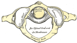
Transverse ligament of the atlas is a thick strong band that stretches across the vertebral foramen of the atlas, holding the dens (odontiod process) of C2 in articulation with the anterior arch of C1. The ligament is attached on either side to a small tubercle on the lateral masses of the atlas. It serves as the seat belt for the odontiod process of C2, allowing just enough movement for the head to move from side-to-side.
 Trauma to this ligament can cause atlanto-axial subluxation. During incidents of trauma to the transverse ligament of the atlas disarticulation between the dens of C2 and the anterior arch, causing spinal cord compression and leading to quadriplegia. This disarticulation could also force the odontiod process of C2 into the medulla of the brainstem, causing death. Symptoms of trauma to the transverse ligament of the atlas is not always noticed by the patient, right away. The vertebral foramen of the atlas is filled with fluid and other tissues still holding the odontiod process in place. Only after an excess of movement could the patient be symptomatic causing quadriplegia or death.
http://en.wikipedia.org/wiki/Transverse_ligament_of_atlas
Herniation of Nucleus Pulposus
 Nucleous Pulposus is a gelatinous substance found in the center of the spinal discs, it is the remnant of the notochord. This core consists of chondrocytes, collagen fibrils, and hyaluronic long chains that attracts water. The nucleus pulposus functions as shock absorbers which distribute hydraulic pressure in all directions within each disc during times of compression, i.e. picking something up. The pulposus is not center in each disc, but instead it lies between the center and posterior aspect of the disc. It is avascular, so it receives nourishment by diffusion from blood vessels at their periphery of the annulus fibrosus and vertebral body. Increased strain or pressure to the nucleus pulposus could cause herniation, the protrusion of pulposus through the wall of the cavity containing it.
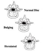
 Herniations of the nucleus pulposus usually occurs posterolaterally, where the annulus fibrosus is thin and does not receive support from either the posterior or the anterior longitudinal ligaments. The posterolateral herniated disc is more likely to be symptomatic because of the proximity of the spinal nerve roots. The acute pain associated with a hernia of the IV disc is a result of the pressure on the longitudinal ligaments and periphery of the annulus fibrosus and from local inflammation caused by chemical irritation by substances form the ruptured nucleus pulposus.
http://en.wikipedia.org/wiki/Nucleus_pulposus
http://en.wikipedia.org/wiki/Nucleus_pulposus
Zygapophysial Trauma
 Zygapophysial Joints are located between the superior and inferior articular processes of adjacent vertebra. This joint is a synovial-plane joint and allow glading movements between the articular processes; the shape and disposition of the articular processes determine the types of movement possible. Range of motion is largely determined by the size of the IV disc relative to that of the vertebral body. A thin joint capsule surrounds the zyapophysial joint; containing the synovial fluid. Each joint is innervated by articular branches that arise from the medial branches of the posterior rami of spinal nerves. The articular branches supply two adjacent joints, so each joint is supplied by two nerves.
Zygapophysial joint trauma or development of osteophytes (bone spurs) effects the adjacent spinal nerves. This can cause pain along the distribution patterns of the dermatomes and spasms in the muscles associated with those spinal nerves. Denervvation of lumber zygpophysial joints is procedure used for treatment of lower back pain. Radiofrequency percutaneous rhizolysis is a neurosurgical procedure that selectively severs problematic nerve roots in the posterior rami spinal nerves.
http://en.wikipedia.org/wiki/Zygapophysial_joint
http://en.wikipedia.org/wiki/Zygapophysial_joint
No comments:
Post a Comment